- Published
Bone Age: A Handy Tool for Pediatric Providers
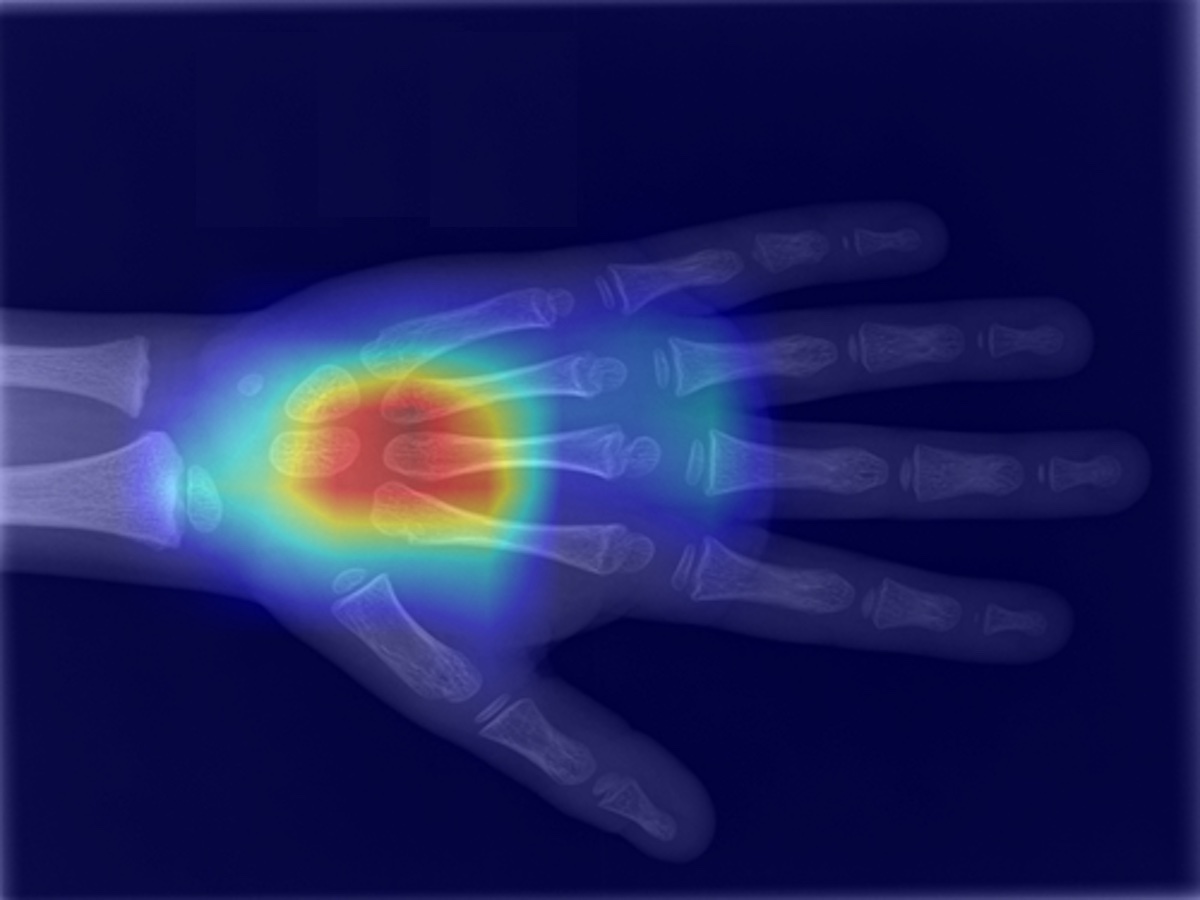
"Heatmap highlights relevant sections" (DALL-E)
For a child's bone age to be considered abnormal, the chronological age must differ from the assigned bone age by more than 2 standard deviations
What's a Bone Age Study?
Bone age refers to how developed a person's skeletal system is. In children, bone age is used as an indicator of physical maturity and can help diagnose issues with growth, hormone disorders, and other medical problems. As someone grows from fetal life to childhood and adolescence and into young adulthood, the bones in their skeleton change size and shape. These changes can be seen through x-rays and other imaging methods. A comparison between the appearance of a patient's bones and a set of standard bone images for a given age can be used to determine the patient's "bone age."
Interpretation
Bone age does not necessarily correspond to an individual's biological or chronological age. For example, someone with stunted growth may have a bone age that is less than their biological age. Similarly, a child growing faster than normal may have a bone age that is older than their chronological age. Most often, a delay or advance in bone age is due to normal variability in growth. However, significant discrepancies between bone age and biological age may indicate an underlying medical condition that requires treatment. Bone age can also be used to predict a child's adult height. Other uses of bone age measurements include diagnosing medical conditions affecting children, such as constitutional growth delay, precocious puberty, thyroid dysfunction, growth hormone deficiency, and other causes of abnormally short or tall stature.
Common techniques
The most common technique for estimating a person's bone age in the United States is to compare an x-ray of the patient's left hand and wrist to a reference atlas containing x-ray images of the left hands of children considered to be representative of how the skeletal structure of the hand appears for the average person at a given age. A paediatric radiologist specially trained in estimating bone age assesses the patient's x-ray for growth, shape, size, and other bone features. The image in the reference atlas that most closely resembles the patient's x-ray is then used to assign a bone age to the patient. Other techniques for estimating bone age exist, including x-ray comparisons of the bones of the knee or elbow to a reference atlas and magnetic resonance imaging approaches.
Clinical significance
To help determine if a child is growing normally, pediatricians assess a patient's bone age. If there is a large difference between a person's bone age and chronological age, it may indicate a growth disorder. For example, if a patient's bone age is less than their chronological age, it suggests a delay in growth, as might be caused by a growth hormone deficiency. If there is too much growth hormone, a child may have a bone age that is older than their chronological age, suggesting they are growing abnormally fast. Since bone age measurements are inherently approximations, they are conventionally reported with a standard deviation, which serves as an estimate of the associated error. For a child's bone age to be considered abnormal, the chronological age must differ from the assigned bone age by more than 2 standard deviations.